Product Introduction
The Kidney-Shaped Lumbar Interbody Fusion Cage is manufactured from PEEK (polyetheretherketone) with integrated tantalum radiopaque markers for intraoperative positioning and postoperative imaging.
Its kidney-shaped anatomical profile facilitates positioning and provides a close fit between the vertebral bodies during lumbar interbody fusion. The cage uses a distraction-compression stabilization principle to provide immediate segmental support, resist rotational and shear forces, and facilitate axial load transfer for intervertebral fusion.
-Features
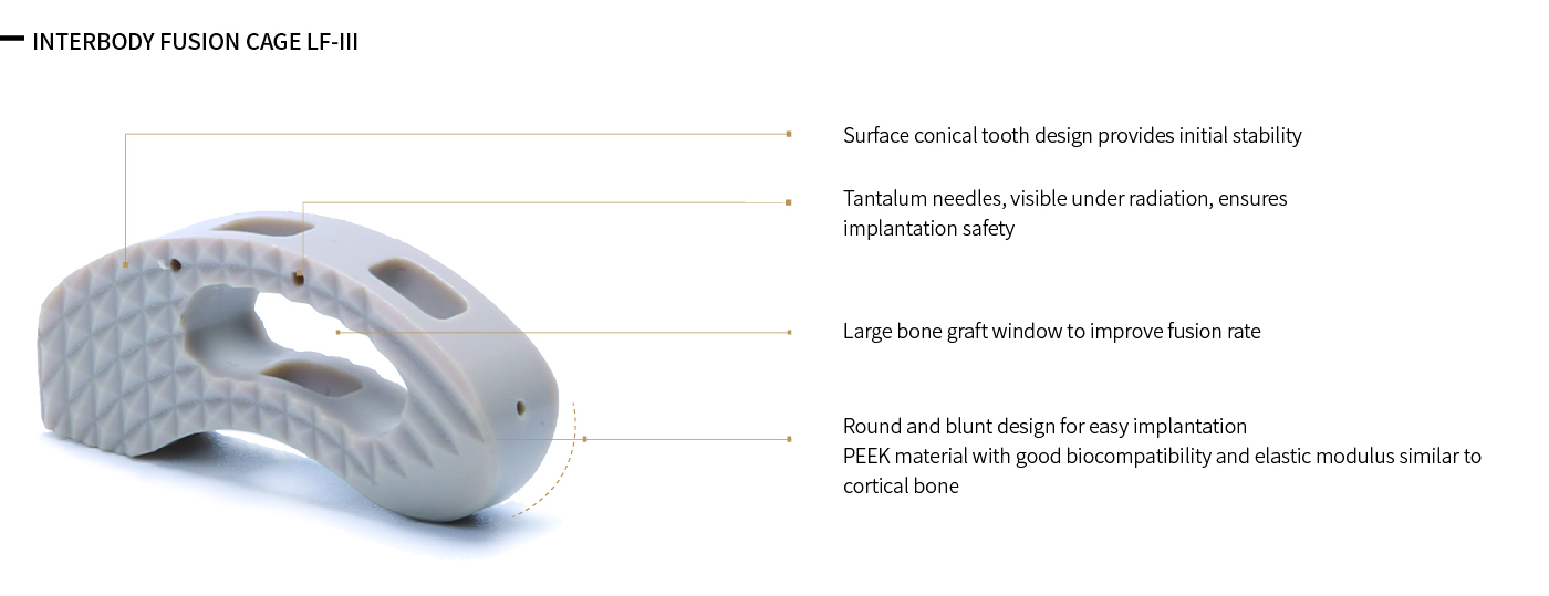
Anatomical Kidney-Shaped Design
• Kidney-shaped profile improves positioning and anatomical conformity.
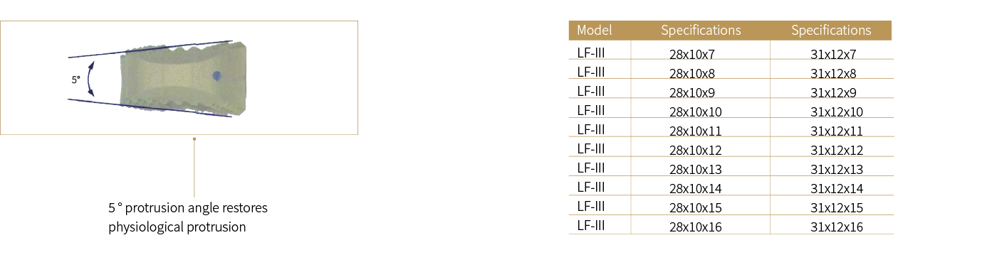
• Wedge-shaped endplates help match vertebral anatomy and maintain sagittal alignment.
Anti-Migration Design
• Pyramidal serrations on the upper and lower surfaces enhance fixation and help prevent cage migration.
Large Graft Window
• The large central graft window accommodates autograft or synthetic bone substitutes to support fusion.
Radiopaque Markers
• Tantalum markers facilitate accurate intraoperative positioning and postoperative imaging.
PEEK Material
• PEEK provides biocompatibility, radiolucency, and bone-like elastic modulus, helping reduce stress shielding and support osseointegration.
Advantage
• Bullet-tip leading edge facilitates smooth insertion.
• Kidney-shaped curvature improves in-body positioning and anatomical fit.
• Wedge-shaped surfaces enhance endplate contact and stability.
• Pyramidal teeth help reduce the risk of postoperative migration.
• Large graft window provides ample space for bone graft material.
• Multiple sizes accommodate different patient anatomies.
• PEEK’s elastic modulus is similar to bone, helping reduce stress shielding and support osseointegration.
• Radiolucent PEEK allows clear postoperative imaging, while tantalum markers provide implant visibility.
Indications
The Kidney-Shaped Lumbar Fusion Cage is indicated for lumbar interbody fusion procedures with supplemental internal fixation.
Typical indications include:
• Degenerative disc disease
• Herniated nucleus pulposus
• Disc space collapse or narrowing
• Revision surgery following failed discectomy or postoperative instability
The device may be used from L2 to S1 based on the surgeon’s assessment and imaging findings.
Specifications