The Self-Stabilizing Cervical Interbody Fusion Cage is designed for anterior cervical interbody fusion and consists of a PEEK (polyetheretherketone) cage, tantalum radiographic markers, and titanium alloy screws. Available in multiple length, width, and height configurations, the cage is positioned between adjacent cervical vertebral bodies and secured with screws to provide immediate postoperative mechanical stability.
The implant incorporates a distraction-compression stabilization concept. Its structural configuration is designed to resist rotational and shear forces while transferring axial loads through the intervertebral space, creating a stable environment for segmental support and bone fusion.
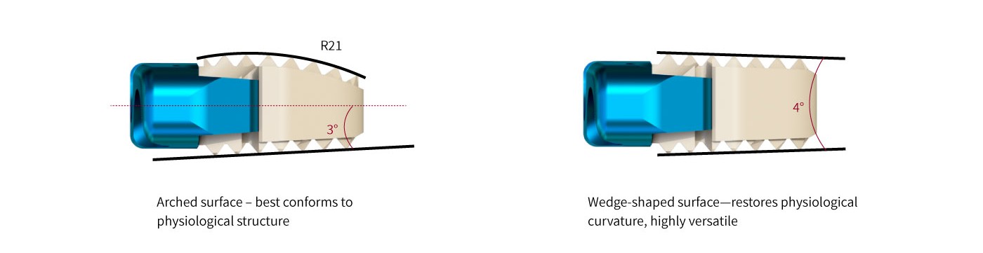
As an interbody spacer, the cage helps restore disc-space height, maintain annular tension and cervical alignment, and support reconstruction of the anterior spinal column. The central graft chamber provides space for bone graft material, supporting the biological fusion process while the implant provides initial mechanical stability.
-Features
Immediate Segmental Stability
The cage is designed to provide immediate postoperative support while helping restore intervertebral disc height and maintain cervical alignment.
Central Graft Chamber
The hollow central structure provides sufficient space for bone graft material, supporting bone growth and fusion across the treated segment.
Zero-Profile Design
The cage is positioned entirely within the intervertebral space, eliminating the need for a conventional anterior cervical plate and helping minimize implant prominence.
Radiographic Visibility
Integrated tantalum markers provide radiographic visibility to assist with intraoperative positioning and postoperative imaging assessment.
PEEK Material
PEEK has an elastic modulus closer to that of natural bone, helping reduce stress shielding and providing a suitable mechanical environment for fusion.
Long-Term Implant Performance
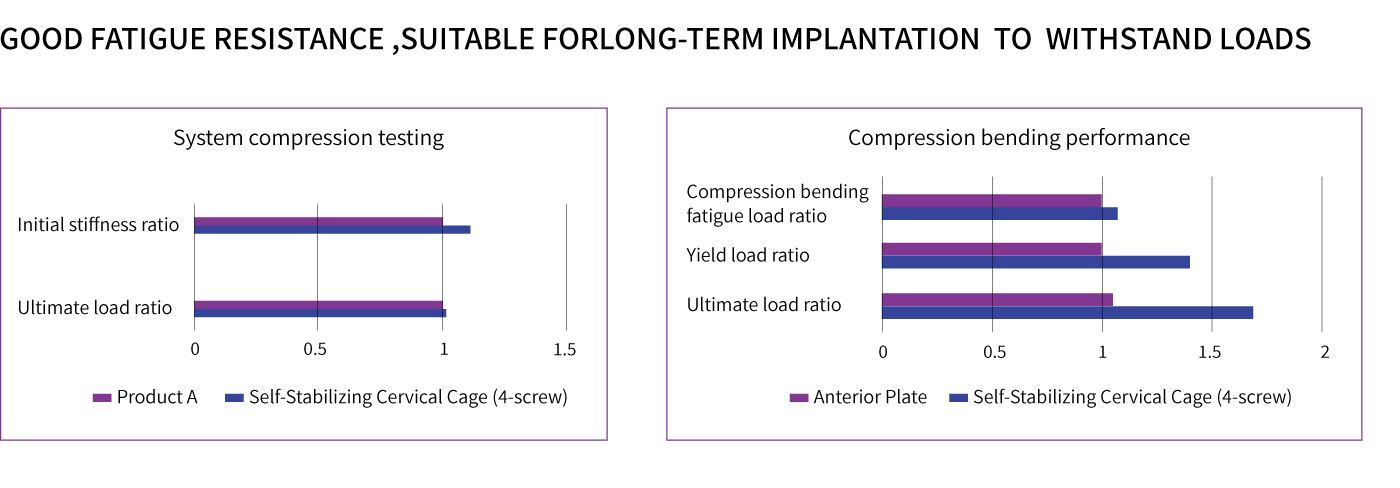
The cage provides fatigue resistance and biocompatibility suitable for long-term implantation.
Imaging Compatibility
The radiolucent PEEK structure allows clear visualization under X-ray imaging, while its MRI compatibility supports postoperative imaging evaluation with reduced artifact.
Secure Screw Fixation
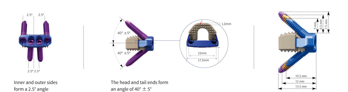
The anti-backout mechanism secures the screws to the cage through snap-ring engagement with the anterior plate structure, helping maintain fixation stability.
-Key Advantages
Multiple Size Options: Various length, width, and height configurations are available to accommodate different anatomical requirements and surgical needs.
Bone-Like Mechanical Characteristics: The PEEK material provides an elastic modulus closer to natural bone, helping minimize stress shielding and support the fusion environment.
Clear Imaging: The radiolucent cage combined with radiopaque tantalum markers provides visibility for implant positioning and postoperative evaluation.
Zero-Profile Fixation: The cage is implanted without a conventional anterior cervical plate, helping minimize anterior implant prominence and the potential for postoperative dysphagia.
Reduced Fixation Components: The screw-based fixation design requires fewer fixation components and may support less invasive surgical approaches with reduced soft-tissue disruption.
Simplified Implantation: The integrated cage and fixation design simplifies the implantation process and may help reduce operative time and surgical trauma.
Material & Design
PEEK Cage: The interbody cage is manufactured from PEEK (polyetheretherketone), providing radiolucency, biocompatibility, and an elastic modulus closer to that of natural bone.
Tantalum Markers: Radiopaque tantalum markers are incorporated into the cage to facilitate implant positioning and postoperative imaging.
Titanium Alloy Screws: Titanium alloy screws provide fixation between the cage and adjacent vertebral bodies.
Zero-Profile Configuration: The implant is designed to remain within the intervertebral space, avoiding the need for a separate anterior cervical plate.
Anti-Backout Mechanism: Snap-ring engagement between the screws and anterior plate structure helps secure the screws and maintain the fixation construct.
The Self-Stabilizing Cervical Interbody Fusion Cage combines a PEEK interbody spacer with integrated screw fixation to provide a compact solution for anterior cervical fusion.
Its zero-profile configuration, central graft chamber, radiopaque markers, and multiple size options provide flexibility for different cervical interbody fusion requirements while supporting both mechanical stabilization and the bone-fusion process.
Application
The device is intended for use in spinal fusion procedures involving:
Cervical Disc Herniation
Degenerative Disc Disease and Intervertebral Space Narrowing
Spinal Instability Following Failed Cervical Disc Surgery
Postoperative Cervical Instability
The device is intended for cases requiring anterior cervical interbody fusion, including patients with or without a previous surgical history.