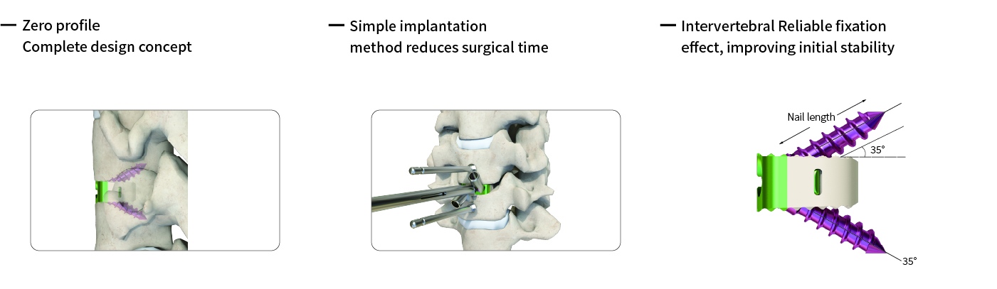
The Self-Stabilizing Cervical Fusion Cage is an interbody spacer manufactured from radiolucent PEEK (polyetheretherketone) and equipped with integrated pure tantalum radiographic markers. The cage is designed for placement between adjacent cervical vertebral bodies and is secured using titanium alloy screws.
The implant is designed to restore intervertebral disc height and support physiological cervical lordosis. Its distraction-compression stabilization concept generates opposing mechanical forces after the disc space is distracted and the implant is secured, helping resist rotational and shear forces and providing early segmental fixation during the fusion process.
Following discectomy, the cage supports reconstruction of the anterior spinal column and transfers axial loads across the treated segment. The implant also helps maintain tension within the annulus and surrounding ligamentous structures. While the cage provides initial mechanical stability, successful fusion ultimately depends on bone growth through and around the implant.
-Features
Immediate Stability and Load Sharing
The hollow-core PEEK structure provides space for bone graft material while helping restore disc height and sagittal alignment.
Serrated teeth on the superior and inferior surfaces engage the vertebral endplates, increasing friction and helping resist implant migration.
Zero-Profile Design
The cage is positioned entirely within the intervertebral space without a protruding anterior plate.
This configuration minimizes contact with surrounding anterior soft tissues and may help reduce the potential for postoperative dysphagia and adjacent-level complications.
Radiographic Markers
Pure tantalum markers are integrated into the cage to facilitate intraoperative positioning and postoperative radiographic evaluation.
The PEEK body is radiolucent, while the tantalum markers provide clear reference points on X-ray and CT imaging.
Bone-Like Elastic Modulus
The elastic modulus of PEEK is closer to that of cancellous bone, helping achieve more balanced load distribution and reduce stress shielding.
The material characteristics, combined with the cage geometry, provide a mechanical environment suitable for bone growth and fusion.
Biocompatibility and Imaging Compatibility
PEEK offers chemical stability and biocompatibility suitable for implant applications.
The radiolucent cage is compatible with postoperative imaging, while the titanium alloy screws and tantalum markers provide mechanical fixation and radiographic reference points.
Secure Locking Mechanism
The cage is secured through screws inserted into a compact fixation block made from TC4 titanium alloy.
A snap-ring anti-backout mechanism between the screws and fixation block is designed to help prevent screw loosening following implantation.
-Key Advantages
Broad Size Selection: Multiple cage footprints and height options provide different length, width, and height combinations for selecting a suitable implant according to patient anatomy and surgical requirements.
Streamlined Implantation: The stand-alone, zero-profile configuration eliminates the need for a separate anterior cervical plate. With a typical two-screw fixation configuration, the system simplifies the implantation process and may reduce operative steps.
Low-Profile Configuration: The flush positioning of the cage within the intervertebral space avoids anterior hardware prominence and may help reduce plate-related complications and postoperative dysphagia associated with conventional plate-and-cage constructs.
Fusion Support: The PEEK cage provides a central graft chamber for autograft or allograft material. Combined with the serrated endplate interface and initial mechanical stability, the design supports the environment required for arthrodesis.
Clear Postoperative Imaging: The radiolucent PEEK body and integrated tantalum markers allow the implant position and fusion progress to be evaluated using X-ray or CT imaging.
Material & Design
PEEK Interbody Cage: The cage is manufactured from radiolucent PEEK (polyetheretherketone), providing biocompatibility and an elastic modulus closer to cancellous bone.
Pure Tantalum Markers: Radiopaque tantalum markers are integrated into the PEEK cage to provide clear reference points during intraoperative and postoperative imaging.
TC4 Titanium Alloy Fixation Block: The lateral fixation block is manufactured from TC4 titanium alloy and accommodates the fixation screws.
Serrated Endplate Interface: Saw-tooth structures on the superior and inferior cage surfaces are designed to engage the vertebral endplates and help resist migration.
Snap-Ring Anti-Backout Mechanism: The screw and fixation block incorporate a snap-ring locking structure designed to help maintain secure screw fixation after implantation.
Why Choose Self-Stabilizing Cervical Fusion Cage
The Self-Stabilizing Cervical Fusion Cage combines a radiolucent PEEK body, integrated tantalum markers, serrated endplate surfaces, and screw-based fixation in a stand-alone zero-profile configuration.
The system is designed to provide initial segmental stability while restoring disc height and providing space for bone graft material. Its multiple size options and compact fixation design offer flexibility for anterior cervical fusion procedures.
Application
The Self-Stabilizing Cervical Fusion Cage is indicated for use in anterior cervical discectomy and fusion (ACDF) procedures from C2 through C7. It is designed to support the vertebral bodies following disc removal and provide stabilization in the following situations:
Degenerative Cervical Disc Disease, including symptomatic disc herniation or degeneration associated with radiculopathy or myelopathy
Disc Space Collapse or Spondylotic Stenosis, including loss of disc height or foraminal narrowing requiring restoration of the intervertebral space
Revision Surgery for failed previous cervical fusion or recurrent disc herniation associated with instability
Postoperative Instability following anterior discectomy where additional stabilization is required
The Self-Stabilizing Cage is designed to provide immediate mechanical support, restore disc height, and support the natural fusion process.